A group of US researchers reported results obtained by coupled micro X-ray fluorescence (µXRF) and X-ray absorption near-edge structure (XANES) for determining the presence of specific sulfur species in coral tissues at high spatial resolution.
Background:
Mapping elements in biological tissues with high spatial resolution is a first step to gain information about tissue structure, organization and function. Especially for essential elements being present in many biomolecules, better information than just the elemental distributioin is needed. Sulfur (S) is omnipresent as an essential element for life, as it is part of organosulfur compounds such as amino acids (cysteine, methionine) and as a cofactor in proteins such as glutathione disulfide. Identifying and mapping the wide range of sulfur species within complex
biological structures presents a challenge for understanding the
distribution of important biomolecules.
In corals, sulfur is present in both the living tissues and nonliving skeletal components as both organic and inorganic phases. Thus, sulfur in corals has been studied from many perspectives, including characterizing sulfate incorporations into aragonitic skeletons as a way to better understand skeletal growth and to deduce the role of organics in biomineralization mechanisms, quantifying dimethylsulfoniopropionate (DMSP) production in the tissues and endosymbionts as an elevated temperature stress signal during coral bleaching events, and measuring S isotopes in carbonate-associated sulfate (CAS) as a paleoproxy.
The new study:
A group of US researchers now has presented a coupled micro X-ray fluorescence (µXRF) and X-ray absorption near-edge structure (XANES) spectroscopy method for determining the presence of specific sulfur species in coral tissues at high spatial resolution. By using multiple energy stacks and principal component analysis (PCA) of a large spectral database, they were able to more accurately identify sulfur species components and distinguish different species and distributions of sulfur formerly unresolved by previous studies.
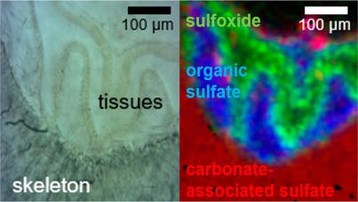
Figure: sulfur species distribution in coral tissue/skeleton obtained by combined XRF/XANES spectroscopy [Reprinted (adapted) with permission from (Anal. Chem., 90 (2018) 12559). Copyright 2018 American Chemical Society]
The results from this combined µXRF/XANES mapping revealed that the coral tissues were dominated by more reduced sulfur species, such as glutathione disulfide, cysteine, and sulfoxide, as well as organic sulfate as represented by chondroitin sulfate. Sulfoxide distributions were visually correlated with the presence of zooxanthellae endosymbionts. Coral skeletons were composed primarily of carbonate-associated sulfate (CAS) along with minor contributions from organic sulfate and a separate inorganic sulfate likely in the form of adsorbed sulfate.
The authors envision that such combined techniques could be applied for mapping metal species with different energy beamlines or using other spatial biological techniques such as fluorescent in situ hybridization probes.
 The original study:
Gabriela A. Farfan, Amy Apprill, Samuel M. Webb, Colleen M. Hansel,
Coupled X‑ray Fluorescence and X‑ray Absorption Spectroscopy for Microscale Imaging and Identification of Sulfur Species within Tissues and Skeletons of Scleractinian Corals, Anal. Chem., 90 (2018) 12559-12566.
DOI: 10.1021/acs.analchem.8b02638
Related studies (newest first)
J.Perrin, C. Rivard, D. Vielzeuf, D. Laporte, C. Fonquernie, A. Ricolleau, M. Cotte, N. Floquet,
The coordination of sulfur in synthetic and biogenic Mg calcites: The red coral case, Geochim. Cosmochim. Acta, 197 (2017) 226-244.
DOI: 10.1016/j.gca.2016.10.017
Yusuke Tamenori, Toshihiro Yoshimura, Nguyen Trong Luan, Hiroshi Hasegawa, Atsushi Suzuki, Hodaka Kawahata, Nozomu Iwasaki,
Identification of the chemical form of sulfur compounds in the Japanese pink coral (Corallium elatius) skeleton using μ-XRF/XAS speciation mapping, J. Struct. Biol., 186/2 (2014) 214-223.
DOI: 10.1016/j.jsb.2014.04.001
Luan Trong Nguyen, Mohammad Azizur Rahman, Teruya Maki, Yusuke
Tamenori, Toshihiro Yoshimura, Atsushi Suzuki, Nozomu Iwasaki, Hiroshi
Hasegawa,
Distribution of trace element in Japanese red coral
Paracorallium japonicum by μ-XRF and sulfur speciation by XANES: Linkage
between trace element distribution and growth ring formation, Geochim. Cosmochim. Acta, 127/15 (2014) 1-9.
DOI: 10.1016/j.gca.2013.11.023
Daniel Vielzeuf, Joaquim Garrabou, Alexander Gagnon, Angèle Ricolleau, Jess Adkins, Detlef Günther, Kathrin Hametner, Jean-Luc Devidal, Eric Reusser, Jonathan Perrin, Nicole Floquet,
Distribution of sulphur and magnesium in the red coral, Chem. Geol., 355 (2013) 13-27.
DOI: 10.1016/j.chemgeo.2013.07.008
Toshihiro Yoshimura, Yusuke Tamenori, Atsushi Suzuki, Rei Nakashima, Nozomu Iwasaki, Hiroshi Hasegawa, Hodaka Kawahata,
Element profile and chemical environment of sulfur in a giant clam shell: Insights from μ-XRF and X-ray absorption near-edge structure, Chem. Geol., 352 (2013) 170-175.
DOI: 10.1016/j.chemgeo.2013.05.035
Alain Manceau, Kathryn L. Nagy,
Quantitative analysis of sulfur functional groups in natural organic matter by XANES spectroscopy, Geochim. Cosmochim. Acta, 99 (2012) 206-223.
DOI: 10.1016/j.gca.2012.09.033
Maggie Cusack, Yannicke Dauphin, Jean-Pierre Cuif, Murielle Salomé, Andy Freer, Huabing Yin,
Micro-XANES mapping of sulphur and its association with magnesium and phosphorus in the shell of the brachiopod, Terebratulina retusa, Chem. Geol., 253/34 (2008) 172-179.
DOI: 10.1016/j.chemgeo.2008.05.007
Yannicke Dauphin, Jean-Pierre Cuif, Murielle Salomé, Jean Susini,
Speciation and distribution of sulfur in a mollusk shell as revealed by in situ maps using X-ray absorption near-edge structure (XANES) spectroscopy at the S K-edge, Am. Mineral., 90/11-12 (2005) 1748-1758.
DOI: 10.2138/am.2005.1640
Jean-Pierre Cuif, Yannicke Dauphin, Jean Doucet, Murielle Salome, Jean Susini,
XANES mapping of organic sulfate in three scleractinian coral skeletons, Geochim. Cosmochim. Acta, 67/1 (2003) 75-83.
DOI: 10.1016/S0016-7037(02)01041-4
Annette Rompel, Roehl M. Cinco, Matthew J. Latimer, Ann E. McDermott, R. D. Guiles, Alexandre Quintanilha, Ronald M. Krauss, Kenneth Sauer, Vittal K. Yachandra, and Melvin P. Klein,
Sulfur K-edge x-ray absorption spectroscopy: A spectroscopic tool to examine the redox state of S-containing metabolites in vivo, Proc. Natl. Acad. Sci., 95/11 (1998) 6122-6127.
DOI: 10.1073/pnas.95.11.6122
K. Xia , F. Weesner, W. F. Bleam, P. A. Helmke, P. R. Bloom and U. L. Skyllberg,
XANES Studies of Oxidation States of Sulfur in Aquatic and Soil Humic Substances, Soil Sci. Soc. Am. J., 62/5 (1998) 1240-1246.
DOI: 10.2136/sssaj1998.03615995006200050014x
Nicholas E. Pingitore Jr., George Meitzner, Karen M. Love,
Identification of sulfate in natural carbonates by x-ray absorption spectroscopy, Geochim. Cosmochim. Acta, 59/12 (1995) 2477-2483.
DOI: 10.1016/0016-7037(95)00142-5
 Related EVISA News (newest first)
last time modified: November 9, 2018